Explore the Fetal Heart
An interactive 3D visualization of fetal cardiac anatomy and prominent defects
Motivation
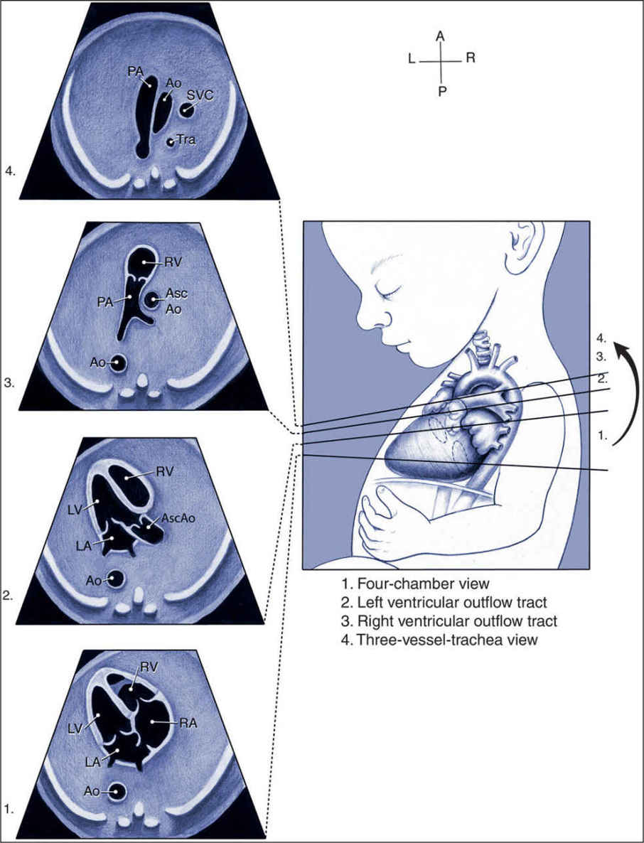
Congenital heart defects (CHDs) are the most common congenital anomalies and represent a leading cause of infant morbidity and mortality. Early detection through fetal echocardiography significantly improves clinical outcomes, however, understanding fetal cardiac anatomy and spatial relationships remains challenging for trainees due to the complexity of intracardiac structures and the limitations of traditional 2D ultrasound instruction.
Current educational tools such as static textbook images, simplified diagrams, and limited-access physical models often fail to adequately convey three-dimensional spatial orientation or dynamic structural relationships. The objective of this project is to develop an interactive, three-dimensional fetal heart model designed specifically for educational and clinical training purposes. The model aims to accurately represent normal fetal cardiac anatomy and common congenital heart defects while allowing users to visualize standard sonographic planes.
By integrating clinically relevant anatomical detail with intuitive user interaction, the design seeks to bridge the gap between 2D imaging and 3D anatomical comprehension. The model is being developed using professional 3D modeling software and optimized for web-based deployment to ensure accessibility, scalability, and ease of use. Emphasis is placed on anatomical accuracy and clear differentiation of intracardiac features relevant to prenatal diagnosis. Prototype development is currently underway, with iterative refinement focused on anatomical modification, accurate representation of common sonographic planes, and usability optimization. Feasibility and educational effectiveness will be evaluated through structured stakeholder surveys to assess anatomical clarity, usability, and perceived instructional value.
Future work includes validation of anatomical accuracy against clinical references and incorporation of additional defect variations based on stakeholder feedback. Ultimately, this design seeks to enhance physician-patient communication and provide a cost-effective alternative to traditional physical teaching models.
Our Sponsor and Acknowledgements
We would like to sincerely thank Dr. Celeste Sheppard for her mentorship, clinical insight, and continued support throughout this project. Her expertise in fetal cardiology has been instrumental in helping us understand the clinical problem and guiding the development of our prototype.
We also extended our gratitude to Dr. Kristen Adams and Dr. Tyrone Porter for their guidance and instruction through the design process. Their mentorship has strengthened our engineering approach and provided valuable direction as we progressed through each phase of the project.
Lastly, we thank the Department of Biomedical Engineering at the University of Texas at Austin for providing the funding, resources, and academic environment necessary to support this work. Without their sponsorship and support, this project would not have been possible.
Our Team
We are a team of four motivated Biomedical Engineering seniors at the University of Texas at Austin with specializations in computation, design, and biologics: Kylie Huynh, Deeya Kaneria, Italia Rodriguez, and Taru Mishra.
Atrial Septal Defect
An Atrial Septal Defect (ASD) is a heart condition that results in a hole between the upper heart chambers from birth. This can increase the strain on the lungs when pumping blood, and harm the direction of bloodflow. Small defects are generally not a cause for concern, however, larger defects may require further surgical intervention.
Ventricular Septal Defect
A Ventricular Septal Defect (VSD) results in the development of a hole in the lower ventricular walls during the womb. This can increase the strain on the lungs when pumping blood, and harm the direction of blood flow because the blood has to flow through the VSD into the lungs. Small defects are generally not a cause for concern, however, larger defects may require surgical intervention.
Tetralogy of Fallot
Tetralogy of Fallot is a condition with four different heart problems: Stenosis, Overriding Aorta, Right Ventricular Hypertrophy, and VSD. This malformation can prevent access to oxygen for the infant and generally requires surgical intervention. It is a rare condition seen to be correlated with Down Syndrome as well.
Patent Ductus Arteriosus
Patent Ductus Arteriosus (PDA) occurs when the opening between the aorta and the pulmonary artery, the Ductus Arteriosus fails to close after birth. This can result in an excess of blood flowing into the lungs and heart, potentially resulting in higher blood pressure, a larger heart, and congestion. Depending on its severity, medicinal or surgical treatment can be employed.